2
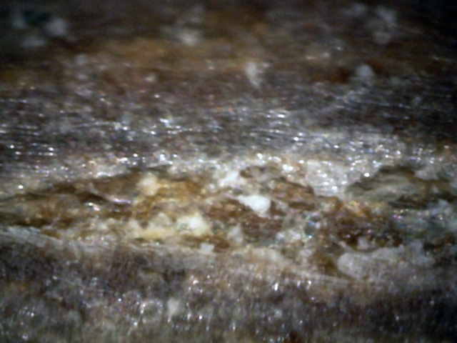
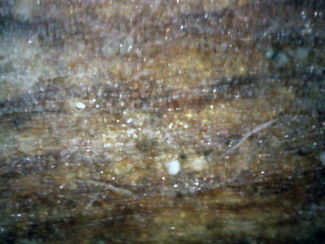
Here there are two spores and two hypha, both next to an area that appears to be
infected with white canker. (400x)
3
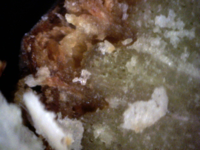
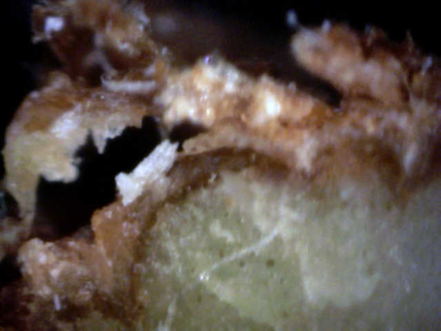
Note the white canker pieces and mangled bark, which also conatins white canker
(although it is more of a yellow-brown in color). (400x)
4
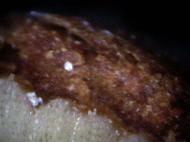
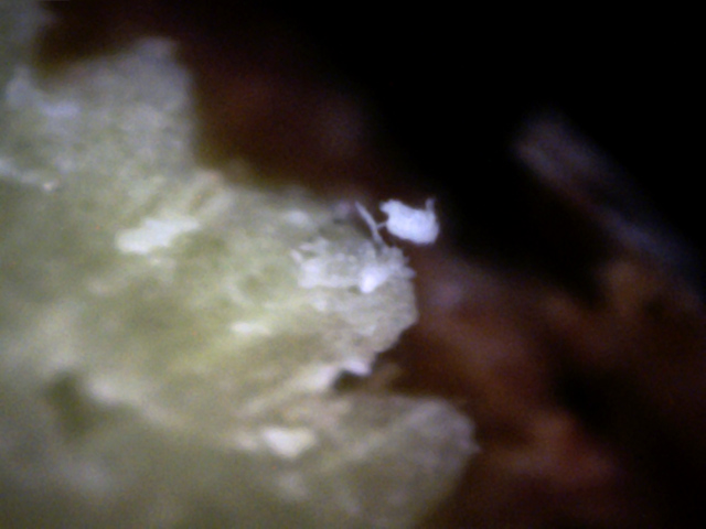
This twig cross-section shows a spore embedded in the bark, and two spores
nestled between the sapwood and bark. Traces of canker material are growing just
to the right of it. (400x)
5
Note the white canker in the sapwood gave rise to a hypha which grew a stalk that generated
another particle of white canker! (400x)
6
The bark here is really mangled due to white canker.
Note the hypha in the sapwood which connects to the blob of white canker
and also to the diffuse area of white canker within the bark.
(400x)
7
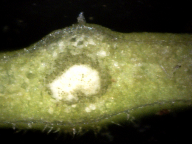
This is a leaf cross-cut perpendicular to the stem.
The white blob in the center is the center vein.
The surrounding gray blobs may be canker.
The small translucent blob on top is probably canker material. (400x)
 ↓
↓
↓
↑
↑
↓
↓
↓
↓
↑
↑
↓
8
In this leaf cross-cut, there is a light gray hypha (black arrows) connecting
three bits of white canker material (red arrows). Hypha semi-transparency makes
them hard to see. (400x)
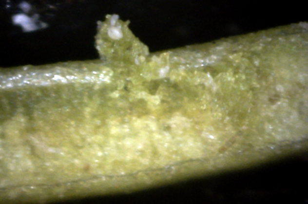
9
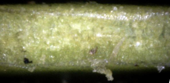
While making this leaf cross-cut, a small piece of leaf surface was ripped up, exposing a canker spore.
This spore apparently had some pieces extending above the leaf surface. (400x)
This shrub, growing in our front yard, almost died from White Canker a few years
ago. Fungal sprays saved it. It had also been sprayed about a month before these
pictures were taken. Therefore, I didn't expect to see too much evidence of
white canker.
The leaves were generally free of white canker. The little that was present is
shown in the following pictures.
